
Dr. Jack Griffin
It’s easy for us dentists to minimize our own significance in overall patient health. We drill holes, we fill holes. We grind, we restore. We clean, we re-clean. Rewarding work but often just becomes routine for some of us. Instead of “just” filling and restoring, we need to understand that we provide therapy. Don’t minimize the effect you can have on a tooth, a smile, even a life.
The effect of removing disease and providing improved oral health is very important for clinicians. It is a satisfying thrill to restore a smile resulting in an increase in happiness and self-confidence for the patient. It is very rewarding when we help a patient chew after years of oral handicap. It is with great gratitude that we get a patient out of pain, save a tooth thought unsavable, or to restore a very compromised occlusion. These are all wonderful services that we provide, life altering in some cases.
Can you help me?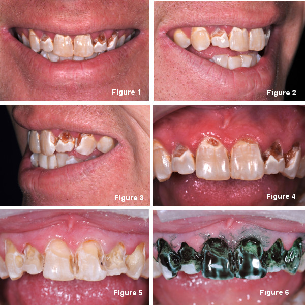
Can we take it a step farther and help save a life? This does not refer to your ACLS training, an epi pen, or basic life support. It instead involves seeing oral disease and investigating the cause. When we see rampant decay, restorations that fail often, or an oral environment that doesn’t fit in with the norm we must try to find out the cause if we want long lasting success.
Of course the patients come to us asking, “Can you help me?” Turn it around. It’s the staff that should be asking “Can you help me?”. It is powerful, non-condemning, and often very revealing. “Can you help me figure out why you are getting this large amount of decay?” “Can you help me determine why your fillings keep breaking?” “Can you help me find out why your teeth are so worn?” Simple yet effective questioning can make a world of difference.
When the doctor examines the patient we often ask about pain, past dental experiences, and goals the patient wants for their smile. Color, shape, and positioning are all very important but not above treating the cause. When we see rampant decay we obviously consider normal etiology such as poor diet, hygiene, and habits. However, the most important question is “Can you help me determine why you have so much decay?”
Substance abuse
The one cause that is often overlooked is substance abuse. In our office we would love to help a patient improve their smile but we need to understand and treat the cause first if want long lasting restorations. With the rise in use of meth, heroin, and other life-destroying drugs we see more destroyed dentitions than we ever have in this practice.
It is easy to typecast the “user” into specific groups but we must be cautioned that abuse is not limited to any race, sex, or socioeconomic group. It has become almost as big a problem with the executive as with the construction worker. These days abuse must be included in the differential diagnosis when determining cause of rampant decay, pre-maturely failing restorations, or oral damage that is beyond the norm.
Drugs like crystal methamphetamine have many chemical and psychological effects on the body. Users suffer oral damage because of very poor diet and little if any hygiene. Teeth grinding and clenching have also been shown to be an effect of the drug. When smoking these drugs chemicals often damage hard and soft tissues leading to red, irritated gingiva and teeth that demineralize and decay rapidly. Salivary flow can also be disrupted contributing to the ease of decay.
Help without condemnation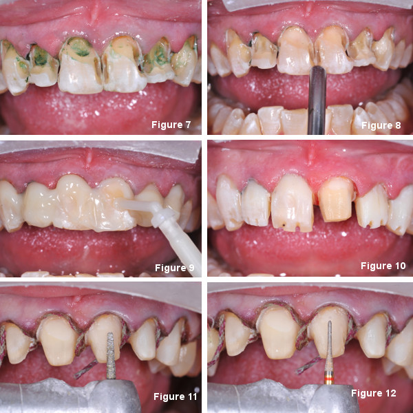
At first meeting the patient and reviewing their concerns, the staff begins by asking the patient for their help in determining the causes of the damage. It is recommended that there is a time in the planning, evaluating process that the staff and the doctor each spend time alone with the patient. It’s amazing how often the patient will say, “I’ve had issues with abuse in the past” to the staff or the doctor when in more of a private setting.
We do not judge, condemn, or criticize. We ask as a health care provider with an interest in the overall well being of the patient. There is no reason to have a great looking restored smile if the inner being is still decayed. We may be the first health care provider that the patient has opened up to about their issues; do not underestimate your potential to help a patient.
What do you do if a patient actually says “I’ve had some issues with medications…”? In a caring manner we simply say “thank you for sharing the information with us, here’s the card of someone we work with to make sure our restorations last as long as possible.” We recommend that each office has business cards or referral slips of someone in your area who is a therapist, counselor, or group who deals with substance. We are providing an avenue for help.
Suspicion does no good if we don’t follow up on it. First we caringly ask and then we have a non-condemning plan for help. Our office policy is that a person be clean and sober one year before elective work; that is the time when there is the greatest chance of relapse. We will get rid of infection, eliminate pain, and other palliative measures before the restorative process actually begins.
Again, don’t be surprised, if your tone and sincerity are right patients will often open up to you. You may save a life.
A case of decay
This case is an example of treatment we provide after substance therapy has concluded for over a year. These cases come to our office 4-5 times per year and once a year we get answers that lead us to offer help. The plan in this case was for lithium disilicate crowns from #5 to #12 with regenerative fillings in all other teeth until definitive restorations could be placed.
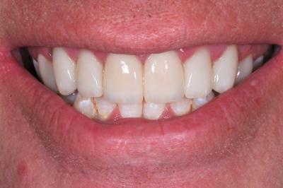
An adult man came to our office with obvious decay. (Fig 1) The consult was for cosmetic treatment and several photos were taken by the staff and put on a large operatory monitor. (Fig 2) We listen to the patient about their desires and ask that they point out areas that they don’t like while looking at their own images on a 35” monitor. Notes are taken as we try to reach a common understanding of how treatment goals are affected by biologic limitations. (Fig 3)
Effects of long-term chemical use often include red, inflamed gingiva, tooth wear, and decay. (Fig 4) Periodontal therapy was completed and then decay was removed with a #4 slow speed round bur and a spoon. (Fig 5) In these cases, thorough decay removal is desired and the teeth should be evaluated with a caries indicator (Sable Seek, Ultradent). (Fig 6) Despite our best attempts, we often leave decay in areas we didn’t evaluate well enough which could lead to prematurely failed restorations. (Fig 7)
Maximizing longevity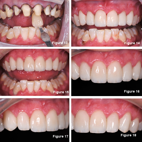
A universal dentin bonding agent was massaged onto the teeth, air thinned, and cured. (Fig 8) A regenerative/bioactive material (Activa, Pulpdent) was then used for the build ups because of its overall strength, ion release, and the potential to re-mineralize and stimulate secondary dentin formation. (Fig 9) These types of materials have the potential to reduce secondary decay by their ion release, re-chargeability, and re-release.
The lithium disilicate preps were begun with 330 bur depth cuts to help insure adequate reduction and postoperative strength. (Fig 10) A tapered diamond was used to do the majority of preparation leaving a rounded shoulder or chamfer margin. (Fig 11) To decrease internal porcelain stresses, the preps are smoothed and corners rounded with a finishing diamond. (Fig 12)
Impressions, alignment guides, and preparation shades are all taken and sent to the lab along with a full series of photos and the goals the patient wants from treatment. (Fig 13) Temporaries were made and the patient given a remineralization–fluoride paste (ReminPro, Voco) to brush with twice a day. The patient was scheduled for insertion and mandibular restorations in 3 weeks.
Insertion and post op care
At insertion the temporaries were removed, the teeth cleaned, and the layered lithium disilicate restorations tried in. (Fig 14) Cementation was done with a high fluoride releasing dual cure giomer resin cement (BeautiCem, Shofu), clean up completed, and the occlusion adjusted. (Fig 15)
Then an impression was taken and a bleach-type tray extending 2 mm onto the gum tissue was made. The instructions were for the patient to place the same remineraliztion-fluoride paste into the tray, in an amount similar to bleach, and sleep with the tray 2 times per week. The purpose is the re-charging of the giomer cement. Brushing with the material at least once a day is to continue as well.
After 6 days the patient returned for a post op visit and the obvious improvement in hard and soft tissue was observed. (Fig 16-18) The patient was re-assured and given praise for the commitment to their overall health and encouraged to continue with our re-care program. We stress the importance of diet and hygiene with the recovered patients while being very positive with them.
The real value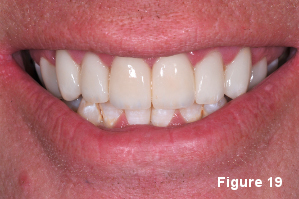
Not only will correcting a defective smile make a patient look good, but the improved self worth may give a patient the confidence to improve their lives in other areas. This may be the self-assurance needed by the patient to restore and maintain themselves and other relationships in their lives. The power of a rehabilitated smile and addressing the destructive causes is immense.
We must not undervalue the potential effect we may have on a patient’s life. Look at your team as important health care providers and with the right questions and treatment you may change a life. You might even save one.
If you have questions about my article or if you would like to send a case, please contact the Pacific Aesthetic Laboratory Group at www.pacificaestheticdentalstudio.com, Gary Vaughn, CDT, CTO (916) 786-6740, or via email [email protected].
The author declared no financial interest in any of the materials mentioned in this article.
The author would like to thank the Pacific Aesthetic Continuum (www.thepac.org) for the principles used in this case and the Pacific Aesthetic Laboratory Group for their excellent case planning and restorations.
Figure legend
Figure 1. Obvious decay and compromised smile.
Figure 2. At the consultation appointment for aesthetic treatment photos are taken to review with the patient
Figure 3. Creating a “shared understanding” of what can be done within biologic limitations
Figure 4. More important than seeing rampant decay is finding out its cause. Poor hygiene, diet, and dental care are usual suspects, but what about substance abuse?
Figure 5. Often chemicals create an environment that fosters aggressive decay that weaken the platform on which the restorations are to be placed.
Figure 6. Caries indicator can increase efficiency in areas of large decay.
Figure 7. Caries indicator is very important as a tool for missed decay and as an aid in knowing when to stop.
Figure 8. Without etch a universal bonding agent was massaged onto the tooth and air thinned.
Figure 9. A regenerative build up material was used for its ion release and strength.
Figure 10. Depth cuts are made with a 330 bur which has a head length of 2mm.
Figure 11. Basic preparations were made with a course tapered diamond.
Figure 12. The preps are smoothed and corners rounded with a finish diamond.
Figure 13. Impressions and all records were taken and sent to the lab.
Figure 14. At the insertion appointment the lithium disilicate crowns were tried in.
Figure 15. Mandibular teeth were resorted with regenerative filling materials and long term plan was for indirect restorations.
Figure 16. Healthy response because of proper diagnosis, personal treatment, and providing an environment where tissues can heal.
Figure 17. The patient was given a bleach type tray with medication to recharge the cement and regenerative filling materials and instructed to wear two nights per week.
Figure 18. Controlling habits and regenerative therapy is the key to long term success.
Figure 19. We in the dental profession must not underestimate the potential we have to alter and perhaps save a life.