

Gary Vaughn, CDT, CTO
As our past participants can attest, the favored reliable technique we employ to simplify the use of a face bow is captured with Panadent’s Kois Dento-Facial Analyzer System. Knowing that a large number of our newsletter subscribers may be unacquainted with this technique, I would like to share not only this modality but also the science that Dr. Kois’ research garnered in developing this increasingly popular technique.
Based on Dr. John C. Kois’ research of an average axis-incisal distance of 100mm, the Kois Dento-Facial Analyzer was developed to simplify the procedures of transferring and mounting study casts for both aesthetics and function. Dr. Kois’ research is substantiated and corroborated by Bonwill’s Equilateral Triangle, Monson’s Spherical Theory (4”=100.12mm), Weinberg studies in 1963 as well as others.
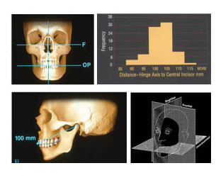 Dr. Kois’ studies were done using different ethnic backgrounds and genders. This bar chart shows the distribution of measurements from the hinge axis to the incisal edge of the maxillary central incisor. As you can see, approximately 80% are within 5 mm of the average 100mm axis-insical distance, which is approximately the same percentages reported in research comparing arbitrary earbows.
Traditionally, dentists are taught to make the incisal canine line parallel to the eyes. If the eyes are slanted, then the teeth would also be made slanted. The dental midline is critical and is always related to the facial midline. Therefore, we need to register the facial midline which dictates the dental midline. Then the occlusal plane will be made perpendicular to the dental midline.
This system registers the steepness and tilts of the occlusal plane related in three planes of space. The horizontal portion of the Analyzer Bow will register an occlusal-horizontal plane of reference. The Vertical Rod will register the facial mid-line for the sagittal plane of reference, and the average axis-incisal distance of 100mm relates to the frontal plane of reference.
Preparation for Registration
Attach Vertical Indicator Rod to Analyzer bow by sliding white attachment disk on the rod into the keyway slot on the Analyzer bow.
Attach disposable Index Tray to Analyzer bow by aligning protruding pins on Index Tray into the holes in the bite fork section of the Analyzer bow. Seat Index Tray all the way flat onto Analyzer bow.
It is best to place 4 Bite-Tabs™ impression compound onto posterior and bicuspid area of the Index Tray. If using registration material other than Bite-Tabs™, first apply an adhesive to occlusal surfaces of Index Tray.
Place Index Tray into a bowl of hot water to temper Bite-Tabs™ impression compound. The compound can be squeezed into a cone shape if more height is required.
Registration
Having the posterior portion of Analyzer Bow down out of occlusion, set the incisal edge of maxillary incisors to the wall or ledge on Index Tray. This registers the incisal point of the average 100 mm axis-incisal distance for function.
Align Vertical Indicator Rod to patient’s facial midline to register the dental midline of the teeth to the frontal plane for esthetics. The Vertical Indicator Rod can be positioned posteriorly in keyway slot of Analyzer Bow to be close to the patient’s nose.
While maintaining incisal contact with the Index Tray and vertical rod alignment to the facial midline, rotate Analyzer Bow up in the posterior until the lateral wings are level to the horizon. This should be done while looking at the front of the patient. The Bioesthetic Level Gauge™ is not required. However, it can be added to verify that the bow is level in the sagittal plane.
These procedures can be more simply done with the patient in a supine position. By doing this, the head is supported by the dental chair head rest. Align the incisal edge to the wall on the Index Tray. You can look at the vertical rod related to the facial midline better from behind the patient. Have the lateral wings hang straight down as you make the registration of the teeth.
You have now captured the steepness and tilts of the occlusal plane in the registration material on the horizontally aligned Index Tray. Remove Index Tray from Analyzer Bow and send to the lab for mounting of study casts. This disposable Index Tray now becomes a permanent bite fork registration record.
If you have questions about my article or if you would like to send a case, please contact the Pacific Aesthetic Laboratory Group at www.pacificaestheticdentalstudio.com, Gary Vaughn, CDT, CTO (916) 786-6740, or via email [email protected].
Reprinted with permission Panadent Corporation.