

Dr. Jack Griffin
Certainly obtaining a great result in a cosmetic case is more than just imagination; it’s a combination of more tangible things. Thorough diagnosis, evaluating patient desires, tempered expectations, prudent tooth preparation, accurate lab records, and proper handling of tissues and materials is critical to success. However, can you see the end before you begin?
Success with cosmetic patients, the most demanding people in our practice, hinges upon what we do BEFORE we lay a bur to the tooth.Two very important and often overlooked aspects of treatment help us visualize the outcome before we crank up the drill – study of the pre-op photos and the lab wax up. Both form the “blueprints” that keep us from going down the wrong road.
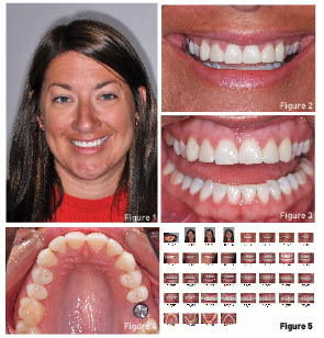 In the case shown here, a young female wants more even color, less square teeth, whiter color and a more feminine look to her teeth. (Fig 1,2,3) She has had an implant placed for an upper molar, porcelain veneers placed on her lateral incisors, and composite bonding done on several teeth. (Fig 4) At the work up appointment a full series of digital photos were taken with a quality camera, flash, and lens system (Nikon D7100, Nikkor 85mm lens, Metz wireless flash). Obviously, great photos are a medical/legal necessity for all practices and you never get a second chance to take pre-operative photos.
Visualization is perhaps as important as any reason to take good images. A full series of images should be standard with views good for case diagnosis, marketing, occlusion, and lab communication. (Fig 5) The chance to sit in a dark room with photos on a large monitor when planning the case is paramount. Images in a place without distraction, even for just a few minutes, can give the clinician a feel for a case that may not be experienced in the business and chaos often seen in the operatories of a busy practice.
Take notes, print and mark up photos, scrutinize, and plan. It’s amazing what I often see on the photos that I didn’t see in the treatment room.
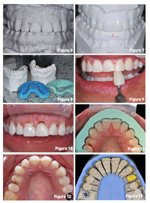 The second part of pre-treatment visualization is models. (Fig 6) Gosh, that sounds boring. On comprehensive cases or where a terrific aesthetic outcome is critical, pretreatment models and photos on a DVD are sent to the lab for a wax up which allows us to see the length of the teeth we have chosen, evaluate the basic smile shape according to patient desires, and have something to show the patient to calm anxiety. (Fig 7) The lab can then create a reduction guide to help insure proper and efficient tooth preparation. (Fig 8)
Photos also document the condition of the teeth before you do any work. Have you ever had a patient forget how bad the teeth looked before you started? Good images document the bite, soft tissues health, and original color which may be important later. (Fig 9) The guide for laser gingivoplasty is a combination of photos and biologic principles. We mark the desired changes on photos with a pen, and after bone considerations, do our shaping. (Fig 10) The images are our reference to remove what we need and preserving what we don’t need altered.
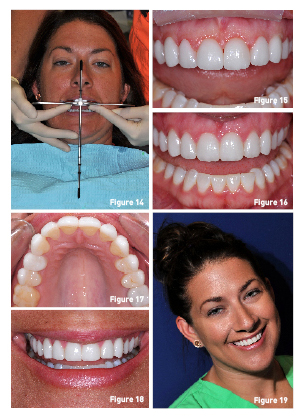 During treatment, the reduction guide is placed and inspected to evaluate tooth reduction. (Fig 11, 12, 13) They can also show the technician different aspects of the case and help identify and even correct problems. Preparation shades help the ceramist chose material opacity and photos of alignment guides, here the Panadent-Kois Analyzer, may show the laboratory if we are off in order for them make that correction for us. (Fig 14)
Confident visualization happens from a thorough exam, quality photos, and accurate lab communication. This minimizes stress for both the clinician and patient. Temporaries made from the wax up are inspected 5-7 days after placement, photos taken, corrections made, and all of this shared with the lab. This results in less stress and minimal issues when the restorations, here lithium disilicate veneers, are tried in the mouth. (Fig 15) After proper cementation, the result is a meeting of the goals of the clinician and desires of the patient. (Fig 16)
The ability to visualize is what many call “an aesthetic eye”. The use of photos and lab wax up can increase your “eye” and keep you on the road to success with minimal stress. (Fig 17) When all of this happens the patient is pleased and the practice will flourish. (Fig 18,19)
If you have questions about my article or if you would like to send a case, please contact the Pacific Aesthetic Laboratory Group at www.pacificaestheticdentalstudio.com, Gary Vaughn, CDT, CTO (916) 786-6740, or via email [email protected].