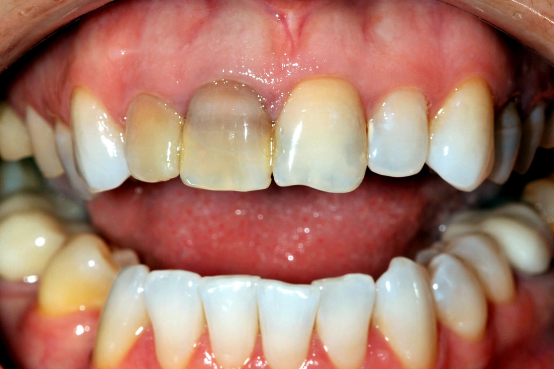
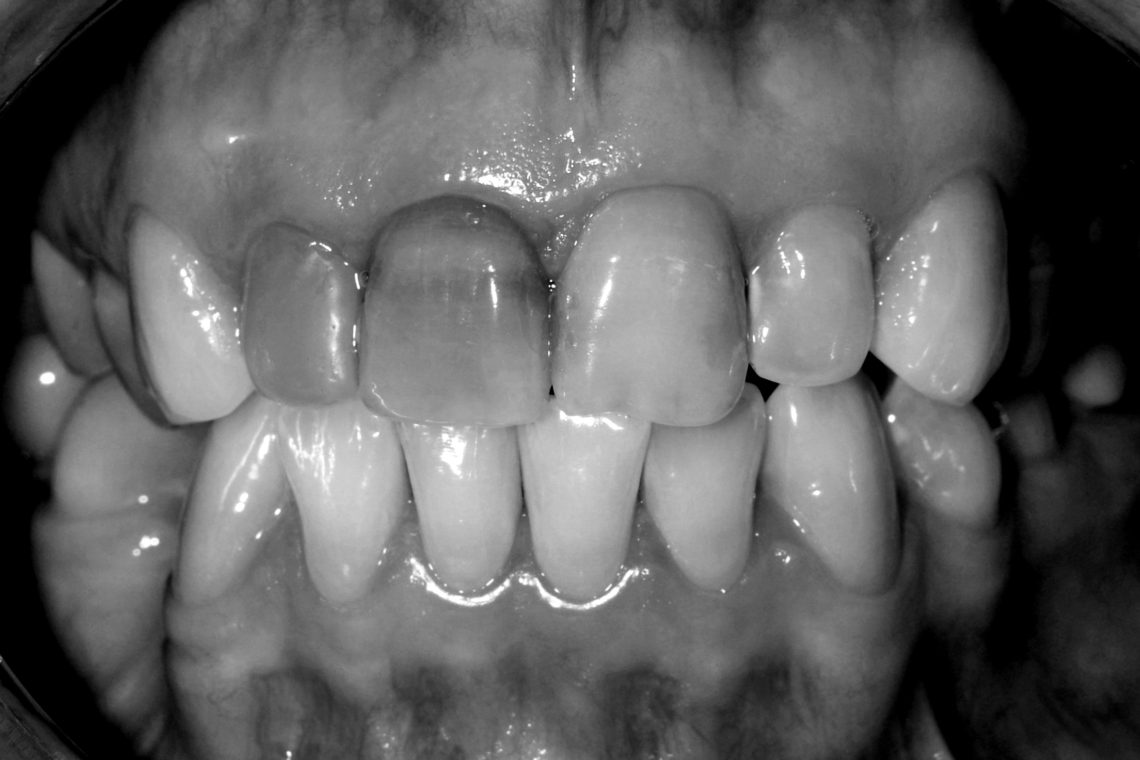
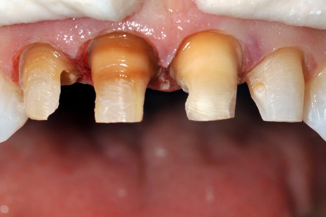
Every ceramist would like to have more room to work…we all would. Every patient wants us to drill less. Particularly when dealing with dark stain and tooth preparation the ceramist says “give me more” reduction, while every patient thinks “please take less” tooth structure away. Years ago we played with varying color cements like “pink opaque” to block grey colors, opaque white to hide metal, and various cement tints to mask colors we didn’t like. Even with try in pastes we were kind of guessing. Today we use materials that have varied opacities which give us more consistent results when masking non-aesthetic discoloration. Two of the more popular materials today are zirconia and lithium disilicate. Zirconia can be very opaque and in a very thin layer has the ability to mask preparation color even in very thin substructure layers. Many zirconia materials in layers of 0.5-1.0mm can block color almost as well as the PFM or Captek crowns only without the metallic color. Lithium disilicate, e.max (Ivoclar), has ingots that come in at least six different opacities. The ceramist takes into consideration the color being covered, the thickness of the prep, and the final color desired in the restoration when choosing the ingot opacity. All of these factors are important and “just more reduction” is merely one factor in the ultimate success of the case. In this case the patient had uneven staining of several teeth, missing lower right molar, and extruded upper right bicuspid. There was interproximal decay or restorations at each anterior contact. She began bleaching in another office and said it made the dark teeth stand out even more than before (fig 1). Sometimes we are “blinded by color” and often don’t notice preoperative variances in value. A black and white photo will often reveal what we don’t see with operatory lights or a bright flash (fig 2).
Figure 1Figure 2Figure 3
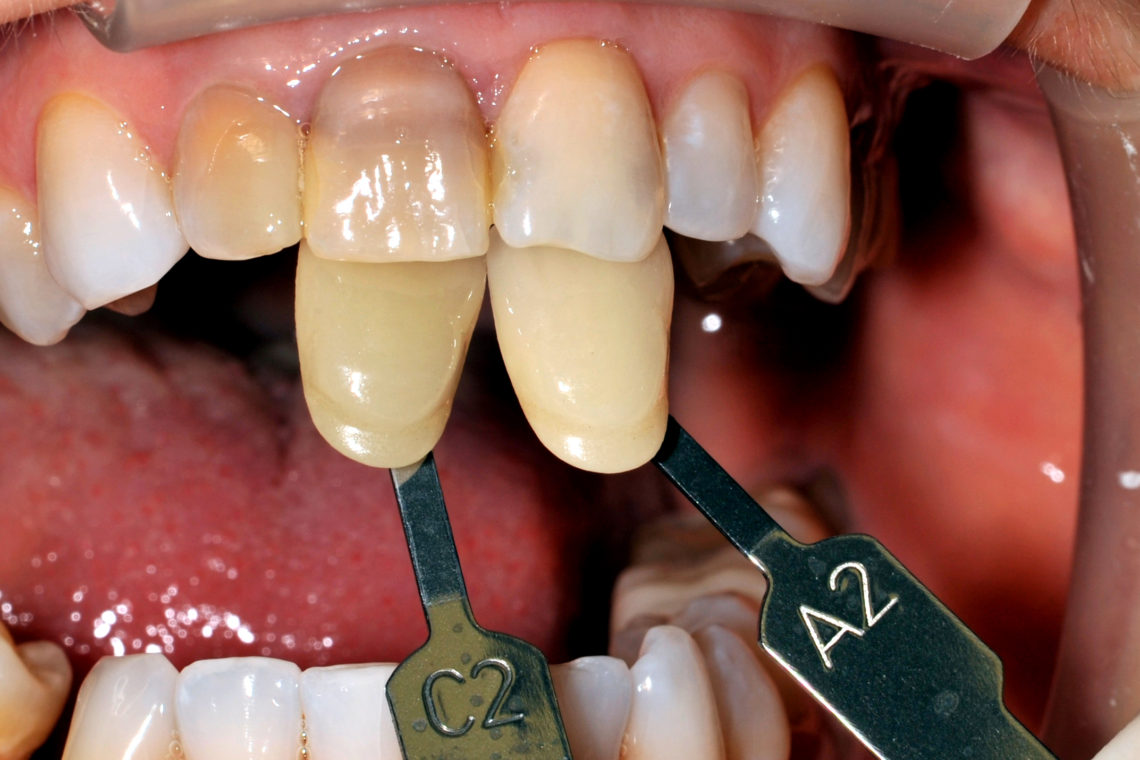
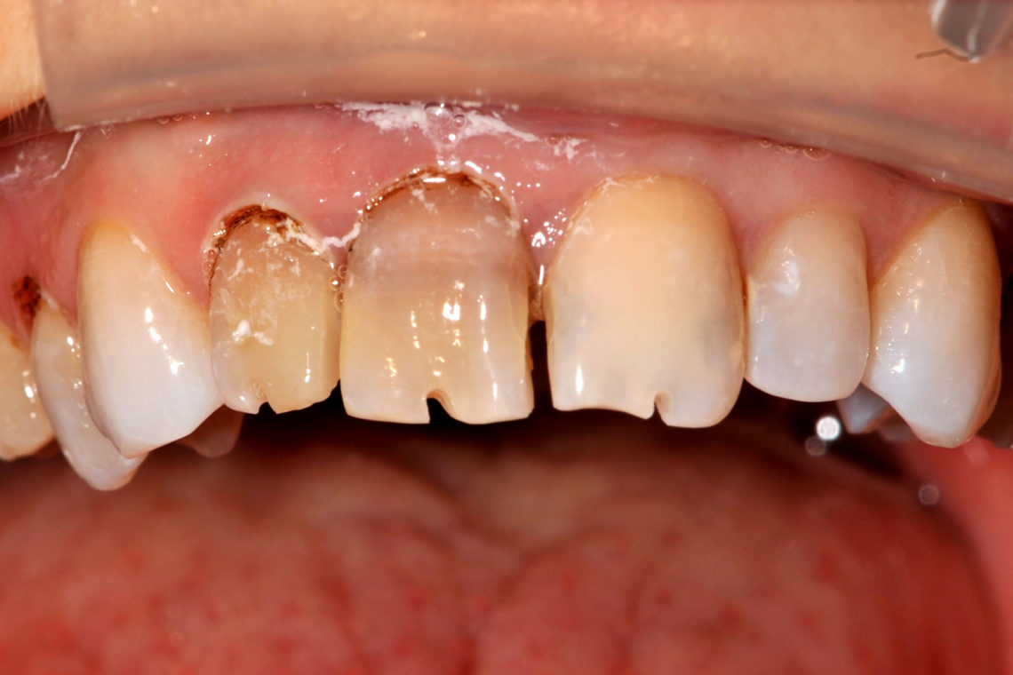
Before prepping any teeth, a full series of photos was taken including preoperative shades, just in case the patient develops “post treatment amnesia” and forgets how they looked before we started (fig 3). In this case, full contour ceramic crowns were to be done from lithium disilicate, e.max (Ivoclar). The only teeth not crowned were the veneers to be done on the cuspids to preserve the cingulum areas. After laser soft tissue re-contouring, depth and interproximal cuts were made with a 330 bur (fig 4).
Figure 4Figure 5
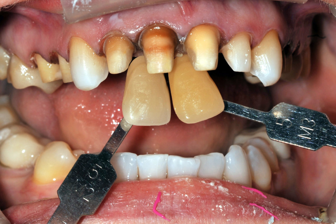
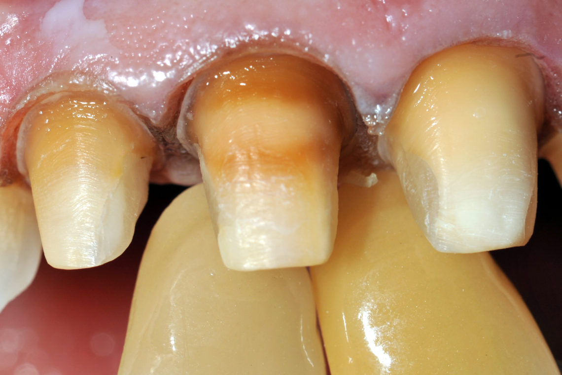
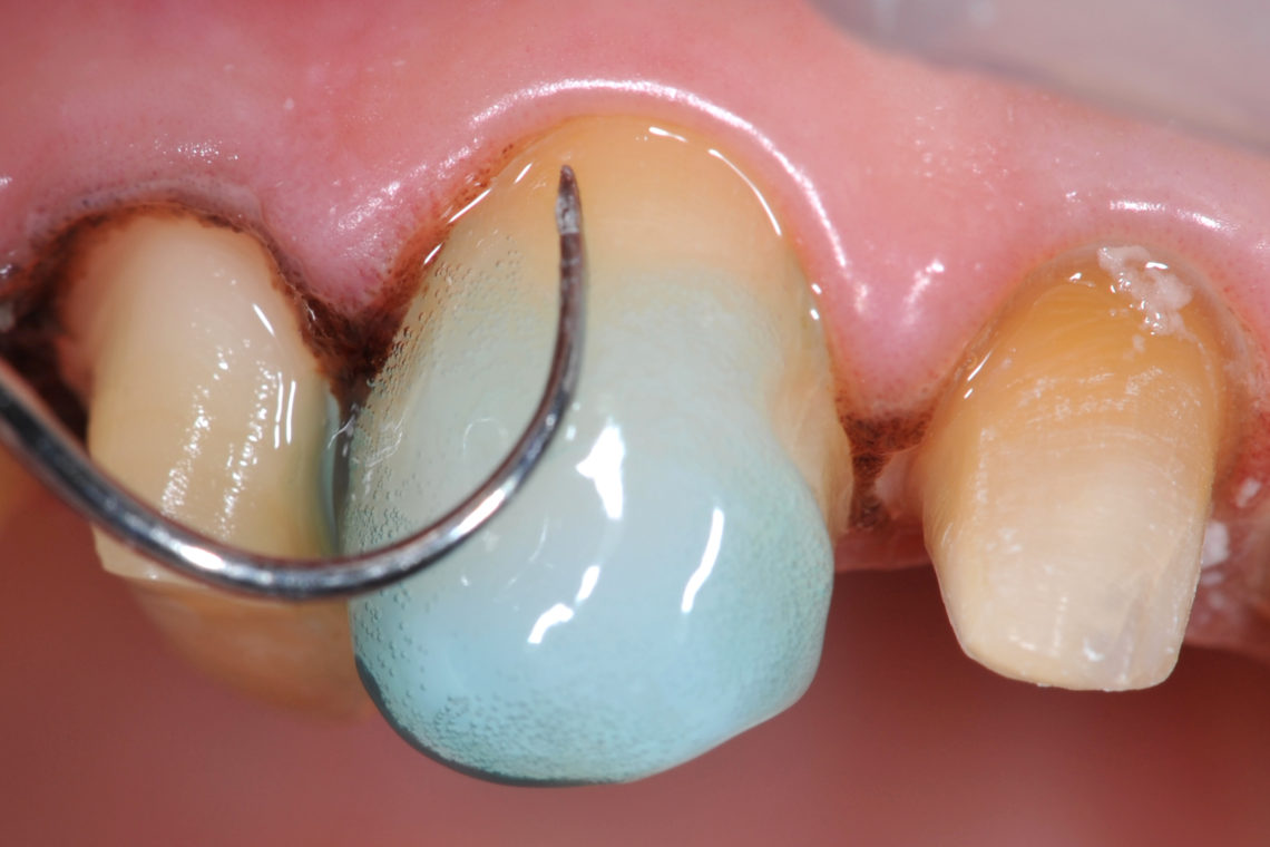
A tapered diamond was then used to complete the crown preparations and decay was removed (fig 5). Often a factor is that the dark color will become more intense as preparation depth is increased. Decayed areas were restored with giomer composite material. The preparation shades were taken to show the lab the intensity of what they need to cover (fig 6). Matching the precise color is not nearly important as communicating the value. With retractors in place to reduce shadows, teeth kept moist, the shade tabs are placed on the lingual and tipped towards the facial so that the middle of the shade tab is on the same plane as the teeth. The first photo shows the entire shade tab and number, the second close up for more precise information (fig 7).
Figure 6Figure 7
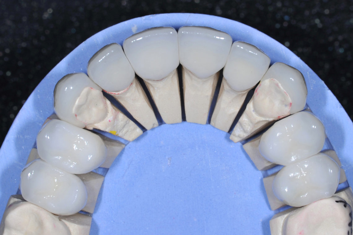
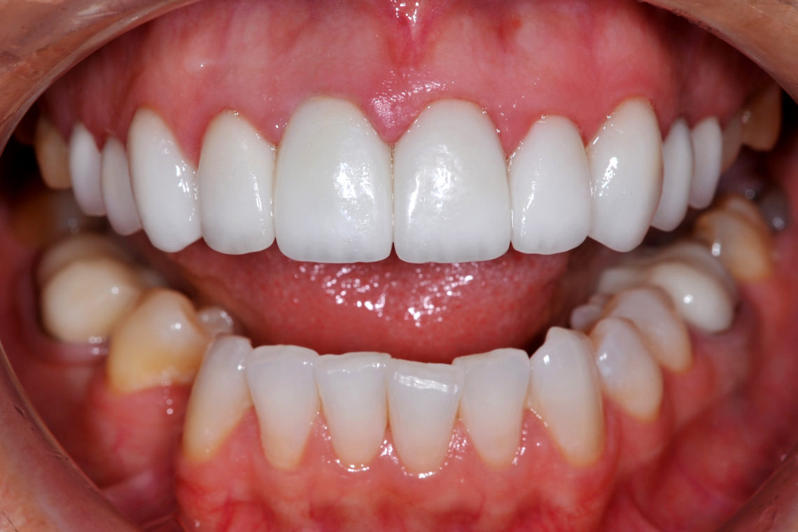
It cannot be stressed enough that how important temporaries are at evaluating the ability to block color. Three to five days after prepping the patient is seen for a post-op visit to evaluate, adjust, and photograph. Along with incisal edge position, shape, and occlusion, the ability of the temporary composite to block color can be communicated to the lab. In this case all restorations were made with an opaque e.max ingot and then layered with character porcelain (fig 8). At insertion, temps were removed and the teeth cleaned. The veneers were placed onto the cuspids with a translucent light cured material after total etch and bonding agent (fig 9). Full contour e.max were all cemented with a translucent dual cure self adhesive resin. Notice the excellent color blockage and the terrific work by the PAC lab to keep hue and chroma consistent between the conservative veneers and the more aggressive full crowns (fig 10).
Figure 8Figure 9Figure 10
Despite compromises in occlusion because of missing lower teeth and inability of the patient to commit to full mouth rehabilitation, the primary concerns of the patient were met. By photographic and written communication with the lab, excellent results can be achieved consistently by understanding the materials being used. If you have questions about my article or if you would like to send a case, please contact the Pacific Aesthetic Laboratory Group at www.pacificaestheticdentalstudio.com, Gary Vaughn, CDT, CTO (916) 786-6740, or via email [email protected]. Article originally printed in PAC TEC newsletter March 2015
By PAC|2020-04-09T16:06:18+00:00March 26th, 2019|Dental|Comments Off on Restorations: Bringing Light to Dark