Dr. Michael Miyasaki
There was a time when doing a complex case meant placing multiple units of porcelain on dark, worn down teeth to the correct aesthetics and occlusion. To accomplish this an understanding of aesthetic preparations, adhesive dentistry and occlusion was about all one needed. Okay, that should be read with some sarcasm; THAT is ALL you needed to know. Today I find that we must be much more knowledgeable in many different areas of dentistry to give our patients the result they seek delivered in the time frame they desire. We need to know how to use lasers, perform endodontic procedures and place implants. In the case presented here our patient presented with these multiple needs, Her main concern was to address those of the maxillary arch and eventually restore the lower. Although the occlusion does not look ideal she was asymptomatic and had apparently healthy joints, muscles and a stable bite. The case shown here required an understanding of: a traumatic oral surgery for the extractions and a knowledge of grafting and immediate implant placement, endodontics in the retreatment of two previously root canaled teeth, periodontics to root plane and scale, prosthodontics (fixed on the maxillary arch and removable on the lower), occlusion, aesthetic design and adhesive dentistry.
We conducted a comprehensive examination noting that there was early periodontitis, multiple teeth with severe decay, no mobility or fremitus, apical lesions on teeth #7 and 10 (which had previous root canal treatment and posts), and multiple missing teeth. Our treatment plan consisted of scaling and root planing the upper arch, preparing teeth #4-6, and #11-13 for bridges, removing the posts from #7 and 10, re-treating the RCT’s on #7 and 10 and placing new bonded posts and cores, extracting #9 with immediate implant placement, extracting teeth (or roots) #1, 5, 12, 14, 15 and grafting for possible implants later, and the delivering of a lower removable prosthetic to replace the missing lower anterior teeth.
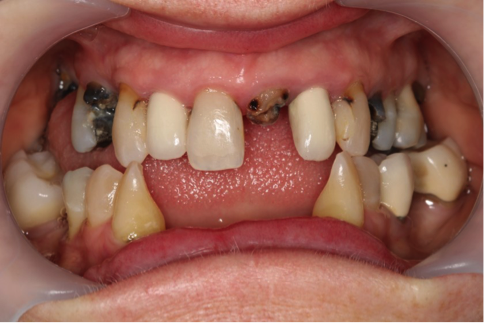
Figure 1
The patient reported that she was anxious about the dental treatment and requested that the maxillary arch be prepared (read as everything being done, but the impression) in one visit. Figure 3 shows the tissues after performing root planning and scaling, retreating teeth #7 and 10 that had the root canals and placing new fiber resin posts and cores, and immediate Megagen implant placement for #9 following extraction. The other decayed teeth were then extracted with their extraction sites grafted during the same visit. This seemed like a lot to accomplish in one visit, but a systematic plan and approach was used. The endodontic system used was the Genius system from Ultradent utilizing a rotary/reciprocating file system. The patient tolerated the treatment well with no significant post-operative sequelae which pleased both the patient and myself.
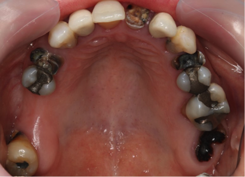
Figure 2
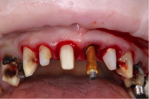
Figure 3
Figure 4 shows the Megagen implant for #9. The ISQ score immediately after placement was a 71 indicating how well the implant adapted to the extraction site giving us very predictable stability for healing.
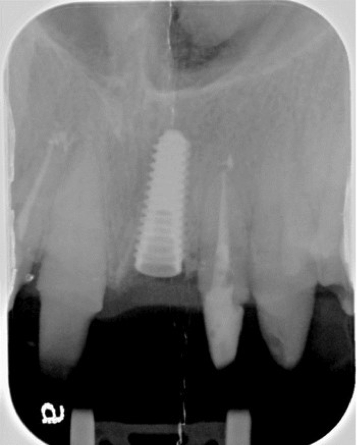
Figure 4
Figure 5 shows the provisional fabricated. The sutures can be seen over #9 and the white streaks on the provisional were from a tint placed over the provisional material to address concerns about the shade. Our patient did not want a Hollywood smile, but something with a natural appearance.
The implant was allowed to heal before final impressions were taken. We used Ultradent’s Gemini dual-wavelength soft tissue diode laser at the impression appointment so we did not have to pack any cord, and we took the final impression using Ultradent’s Thermo Clone VPS impression material because of its accuracy and dependability with multiple unit cases.
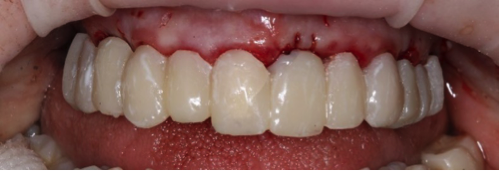
Figure 5
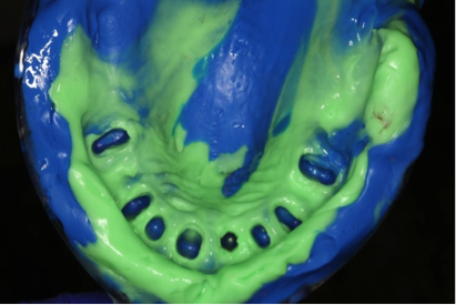
Figure 6
Corr Dental Designs and Todd Fong, CDT, did an amazing job with all the units going right into place at the cementation appointment without any adjustment. Figures 7 and 8 show the pre-operative condition and the immediate results. Our patient was very happy with the aesthetics and the case should look even better today after a few weeks of healing.
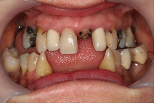
Figure 7
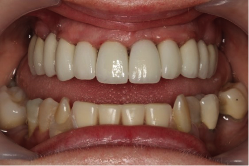
Figure 8
Today’s clinician needs to be a constant student, and be open to learning so that they can offer their patients treatment options that best fit their aesthetic and functional desires and needs. Come join me at www.p-bdi.com, and take part in the education offered by PAC. You will learn more to help your patients, grow your practice and increase your professional satisfaction.
If you have questions about my article or if you would like to send a case, please contact the Pacific Aesthetic Laboratory Group at www.pacificaestheticdentalstudio.com, Gary Vaughn, CDT, CTO (916) 786-6740, or via email gvaughn@thePAC.org.
Figure 1. Pre-op maxillary arch-frontal view.
Figure 2. Pre-op maxillary arch-occlusal view.
Figure 3. Arch prepared and an implant was placed for #9.
Figure 4. Implant and root canals completed.
Figure 5. Provisionals.
Figure 6. Showing the impression using Ultradent’s Thermo Clone VPS material.
Figures 7 and 8. Pre-op and Final case immediately after cementation.
